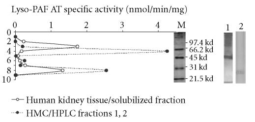
Figure 7.
Native-PAGE electrophoresis: Lyso-PAF AT specific activities on native PAGE electrophoresis of solubilized fractions of human kidney tissue (-∘-) and active HPLC fractions 1, 2 of mesangial cells (-•-). The Log MW of standard proteins versus mobility was linear. Lines M, 1, and 2 represent the part of the gel stained by Coomassie Blue from molecular markers, human kidney tissue, and HMC, respectively.

