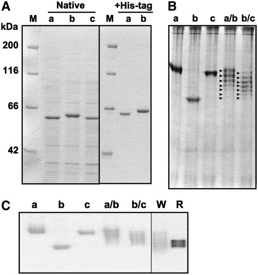
Figure 3.
SDS- and native-PAGE of the wheat and rye glucosidases. A, The glucosidase monomers were analyzed by SDS-PAGE. For the native (without His-tag) glucosidases, the crude E. coli cell extracts were directly subjected to SDS-PAGE. The His-tagged glucosidases were electrophoresed after purification by affinity chromatography on a nickel-charged column. B, The cell extracts were subjected to native-PAGE (on an 8% separating gel for 4 h) and stained with Coomassie Brilliant Blue. The arrowheads indicate the seven types of glucosidase hexamers expressed in coexpression lines. C, The bands with β-glucosidase activity were detected by activity staining. The crude extracts of E. coli cells and 48-h-old shoots were separated under nondenaturing conditions. In each segment, M, a, b, c, a/b, b/c, W, and R indicate marker proteins, TaGlu1a, TaGlu1b, TaGlu1c, coexpressed TaGlu1a and TaGlu1b, coexpressed TaGlu1b and TaGlu1c, wheat shoots, and rye shoots, respectively.

