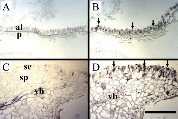
Figure 5.
Cellular localization of hexose transporter mRNAs in germinating wheat seeds. Photomicrographs of longitudinal sections of 3-DAI paraffin-embedded germinating wheat seeds treated with sense (A and C) and antisense (B and D) probes from a highly conserved hexose transporter sequence. Probe binding was visualized using an alkaline phosphatase reaction yielding a blue-purple product. A and B, Longitudinal sections through the aleurone and pericarp tissues. Note the signal of the hexose transporter transcript (arrows) is localized to the aleurone cells (arrows). C and D, Longitudinal sections through the scutellum region showing the hexose transcript signal (arrows) localized to the columnar-shaped epidermal cells and the ground cells of the scutellum. Note the absence of signal from the scutellum vascular bundle. al, Aleurone; p, pericarp; se, scutellum epidermis; sp, scutellum parenchyma; vb, scutellum vascular bundle. Scale bar = 200 μm.

