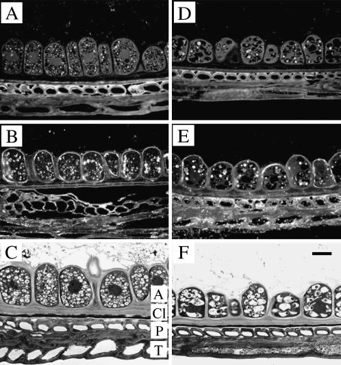
Figure 6.
Cellular localization of TaSUT1 proteins in the aleurone layer of germinating wheat seeds. A and D, Immunofluorescent images of aleurone sections from 3-DAI (A) and 7-DAI (D) seeds treated with preimmune serum and secondary antibody. Note the strong autofluorescence of protein bodies in the aleurone cells and walls of the sclerenchyma cells located in the pericarp. B and E, Immunofluorescent images of aleurone sections from 3-DAI (B) and 7-DAI (E) seeds treated with the SUT1 antibody, detected as a fluorescent signal emitted by FITC-conjugated anti-rabbit secondary antibody. Note that immunofluorescence is restricted to the plasma membranes of aleurone cells. C and F, Bright-field micrographs stained with toluidine blue illustrating the degradation of aleurone cell structure, including characteristic changes in vacuole composition. A, Aleurone; Cl, cuticle layer; P, pericarp; T, testa. Scale bar = 25 μm.

