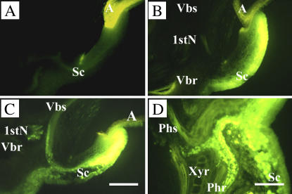
Figure 8.
Symplasmic movement of CF in the embryo of 3-DAI wheat seedlings. The surface of the scutellum epidermis was exposed to CFDA solution for 3 min, washed exhaustively, and then examined by fluorescent microscopy. A, Autofluorescence from scutellum and aleurone, detected with the same microscopic setting as used in B to D. B, Distribution of CF in embryonic tissues after 3-min exposure and washout. C and D, Distribution of CF after chasing for 4 h. A, Aleurone; 1stN, first node; Phr and Phs, phloem directed to root and shoot; Sc, scutellum; Vbr and Vbs, vascular trace entering root and shoot; Xyr, xylem directed to root. Scale bar = 50 μm.

