Figure 5.
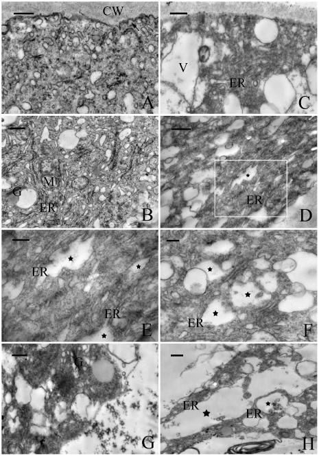
Electron micrographs of pollen tubes of P. wilsonii cultured in standard medium for 24 h (A and B) or treated with 40 μm MG132 for 20 h (C–G) or 24 h (H). A, The apical region of a control tube showing vesicle-rich zone. Note that some of these vesicles are fusing with the plasma membrane (indicated by arrow). B, The subapical region of a control tube, showing abundant organelles including ER, mitochondria (M), and Golgi stacks (G). C, The apical region of a treated pollen tube, showing dramatic decline in vesicles and appearance of vacuoles (V) and other organelles in this zone. D to H, The subapical region of treated tubes, showing MG132 induced ER dilatation (indicated by stars) and cellular vacuolization. Figure E represents magnified view of boxed area in Figure D (indicated by box). Bar = 0.5 μm for A to D and H and 0.2 μm for E to G.

