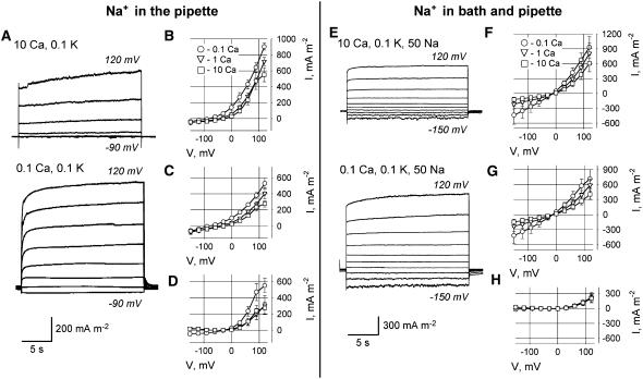
Figure 7.
Whole-cell plasma membrane currents (protoplasts derived from Arabidopsis mature root epidermis) with 50 mm Na+ present in the patch pipette (A–D), and when 50 mm Na+ was present both in the bath and PS (E–H). A, Typical currents (recorded from the same protoplast) in 10 mm (top) and 0.1 mm (bottom) extracellular Ca2+ for Na+ present in PS only. B to D, Mean ± se I-V relationships (n = 4–5) for total (B), instantaneous (C), and time-dependent (D) currents measured at 0.1, 1, and 10 mm extracellular Ca2+. E, Typical currents (recorded from the same protoplast) in 10 mm (top) and 0.1 mm (bottom) extracellular Ca2+ for symmetrical 50 mm Na+ conditions. F to H, Mean ± se I-V relationships (n = 4–5) for total (F), instantaneous (G), and time-dependent (H) currents measured at 0.1, 1, and 10 mm extracellular Ca2+. Time-dependent currents were calculated as described in “Materials and Methods.” PS contained 50 mm potassium gluconate, 30 mm KCl, and 50 mm sodium gluconate. Extracellular K+ was 0.1 mm. All concentrations are given in mm.

