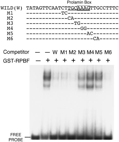
Figure 5.
EMSA of RPBF protein with a P box in the GluB-1 promoter. Nucleotide sequences of oligonucleotides used as probe and competitors are depicted. WILD, 29 bp sequence containing a P box from the GluB-1 promoter (−152 to −123); M1 to M6, 29 bp sequence with successive dinucleotide mutations. The P-box and AAAG motif are underlined and boxed, respectively. The GST-RPBF fusion protein was used for EMSA with the 29 bp sequence containing the P box. Competitors were added in 100-fold molar excess. Lane 1, No protein; lane 2, no competitor; lanes 3 to 9, with competitors (wild type [W] and M1 to M6).

