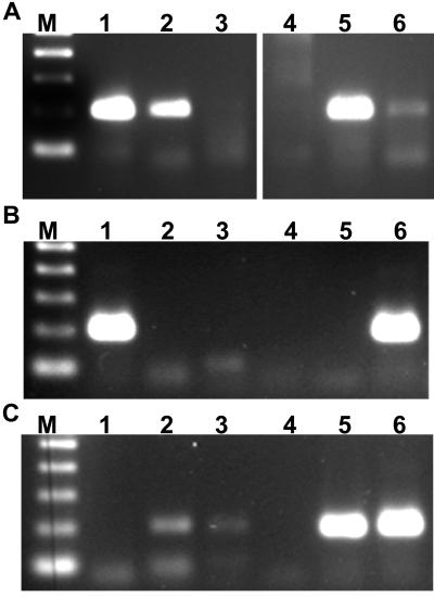
FIG. 6.
Detection of E. canis in experimentally infected ticks. R. sanguineus adult male ticks were infected as nymphs and incubated at RT or 37°C prior to aseptic bisection, followed by digestion of individual tick halves with proteinase K in the presence of nonionic detergents. (A) Pooled digests of ticks subjected to proteinase K digestion alone (lanes 2 and 5) or to DNA purification (lanes 3 and 6). Lanes 1 and 4 represent infected canine buffy coat DNA and a template-free control, respectively. (B) Individual ticks incubated at RT (lanes 2 to 6) Lane 1 represents infected canine buffy coat DNA as a positive control. (C) Individual ticks (lanes 2 to 6) incubated at 37°C prior to bisection and proteinase K digestion. Lane 1 represents a template-free control.

