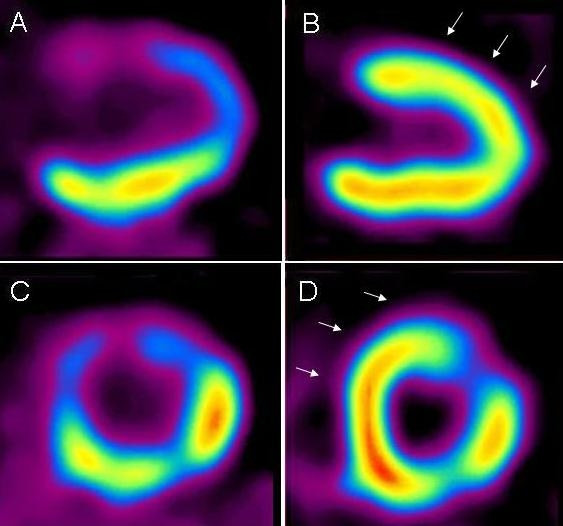
Figure 4.

Vertical long axis (A, B) and short axis (C, D) 18F-FDG-PET images of patient with myocardial infarction. Non-transmural defect at apex and anterior wall before surgical therapy (A, C). Three months after cell transplantation and CABG, anterior wall and apex showed increased viability (arrows) in infracted area (B, D). EF changed from 31% to 60%.
