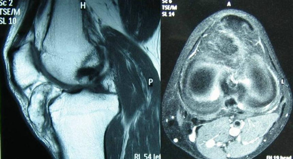
Figure 2.

A: T1-weighted image: slight joint effusion. A mass located in the anterior portion of the space joint is evident. A pedicle is seen, but there was no continuity with the tibial plateau. The mass seems to contain some chondral components. B: T2-weighted image at the level of the tibial plateau shows intralesional bone formation (hypointense areas).
