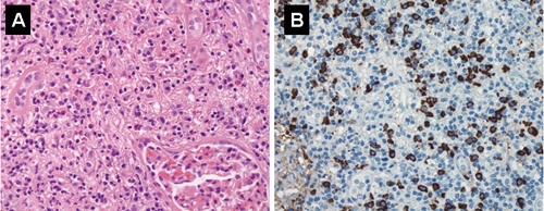
Fig. 3: A: Renal lesion showing extensive lymphoplasmacytic inflammatory infiltrate with scattered eosinophils; note interstitial fibrosis and almost complete loss of tubules (hematoxylin–eosin stain, original magnification × 400). B: Many inflammatory cells stain positive for IgG4 (immunoperoxidase, original magnification × 400).
