
Fig. 5. Complex formation between BiP and J-MTJ1.
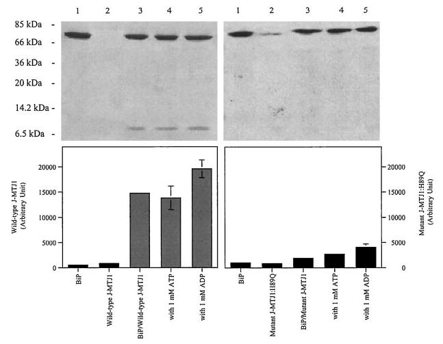
Interaction of His6-BiP with J-MTJ1 (left panels) or J-MTJ1: H89Q (right panels) was assayed as described under “Experimental Procedures.” Top panels, Coomassie-stained 15% SDS-PAGE. Lane 1, His6-BiP; lane 2; cleaved J-MTJ1 (or, right panel, mutant J-MTJ1:H89Q); lane 3, His6-BiP with wild-type J-MTJ1 (or, right panel, mutant J-MTJ1:H89Q); lane 4, His6-BiP with wild-type (or, right panel, mutant J-MTJ1:H89Q) in the presence of 1 mm ATP; lane 5, His6-BiP with wild-type (or, right panel, mutant J-MTJ1:H89Q) in the presence of 1 mm ADP. Bottom panel, densitometry scanning of the SDS-PAGE. The y axis represents the average of three independent experiments.