
Fig. 6.
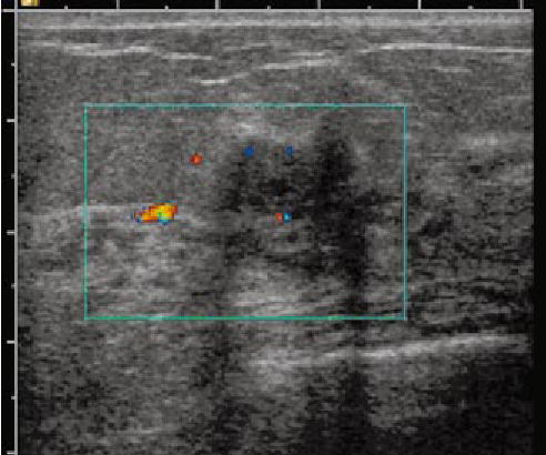
Ultrasound gray-scale image with Doppler superimposed. The lesion was located at the 3 o’clock position of the left breast of a 45-year-old patient and was considered as highly suspicious. The ultrasound revealed irregular shape and posterior shadowing with blood flow seen at several periphery sites of the lesion.