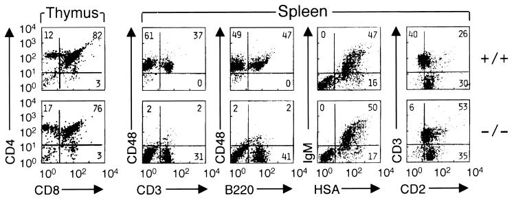
Figure 2.
Lymphoid development in CD48-deficient mice. Thymocytes and splenocytes were purified from CD48+/+ mice (upper row) and from CD48−/− mice (lower row) and were analyzed by dual color immunofluorescence and flow cytometry. All samples were analyzed on a FACScan after compensation. Five-thousand cells were analyzed per sample. Antigens displayed on the y axes were stained with PE-conjugated mAbs whereas antigens on the x axes were stained with fluorescein isothiocyanate-conjugated mAbs.

