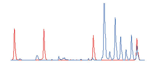
Figure 1.

An example of an ATAC-PCR electropherogram. The height of each peak represents fluorescence intensity. Blue, PCR product signals; red, size marker signals. Starting from the left, the marker sizes are 35, 50, 75 and 100 bases. The seven blue peaks on the right correspond to ATAC-PCR products. From the left, the first peak corresponds to 10 equivalents of control cDNA, the second corresponds to two equivalents of control cDNA, and the last five peaks correspond to five equivalents of sample cDNA.
