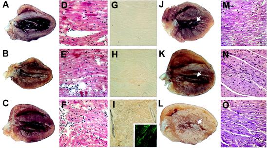
Figure 1.
Representative gross anatomy, histology, and immunohistology of normal mouse hearts compared with hearts from transgenic mice expressing the normal human ELC (human control) or mutant human ELC (mutant human) at 3 months and at 1 year of age. (A–C) Hearts from normal (A), human control (B), and human mutant (C) mice, respectively, at 3 months of age are similar. (D–F) The morphology in H- & E-stained sections of the three hearts shown in A–C is normal. (×50.) (G–I) Immunoreactivity after staining with polyclonal antibody against mutant ELC is absent in normal hearts (G), minimal in human control hearts (H), and strong in the human mutant hearts (I) HR peroxidase method was used. (×50.) (I, Inset) Localization of reactivity in myofibrills by using a fluorescein isothiocyanate-conjugated (green) secondary antibody. (×150.) (J–L) Hearts from three different normal (J), human control (K), and human mutant (L) mice at 1 year. Solid arrows in J and K show normal left ventricular cavity. Arrow in L shows hypertrophied papillary muscle obstructing the middle of the mostly obliterated left ventricular cavity. (M–O) H- and E-stained sections of normal (M), human control (N), and human mutant (O) hearts at 1 year. M and N are normal; O shows myocyte hypertrophy without disarray. (×50.)

