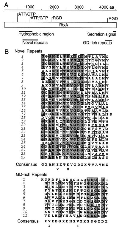
Figure 4.
The potential structural domains and motifs in V. cholerae RtxA and sequence comparison to other RTX toxins. (A) The predicted RtxA polypeptide is represented schematically by an open box with its length in amino acid residues indicated on the line above. The positions of potential ATP/GTP-binding domains and the two RGD motifs are indicated. Larger putative structual domains are identified by solid bars. (B) Sequence alignments of repeats from the hydrophobic region (amino acids 781-1332) and repeats from the GD-rich region (amino acids 4165–4362) of V. cholerae RtxA.

