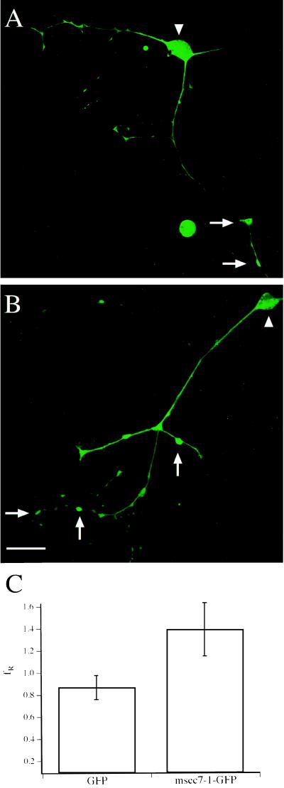
Figure 2.
msec7-1 is located predominantly to presynaptic regions of Xenopus nerve cells. Shown is the fluorescence pattern of spinal cord neurons injected with a mixture of msec7-1 and GFP mRNA (A) or with msec7-1-GFP mRNA (B). Note that the postsynaptic muscle cells in these examples are not visible because of their lack of GFP fluorescence. Arrows, varicosities; arrowheads, cell bodies. (Bar = 50 μm.) (C) Quantitative analysis of fluorescence intensities. The ratio of varicosity to soma fluorescence (fR) in GFP and msec7-1-GFP containing neurons was 0.87 ± 0.11 (n = 8) and 1.39 ± 0.24 (n = 8), respectively. Error bars are given as SEM.

