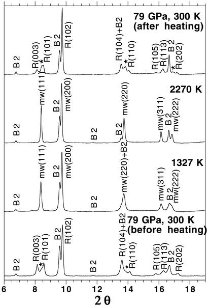
Figure 2.
Representative angle-dispersive x-ray diffraction patterns of (Mg0.25,Fe0.75)O at ≈79 GPa in a laser-heated DAC. A monochromatic beam (wavelength = 0.3311 Å) was used as the x-ray source. The backgrounds were subtracted with PEAKFIT 4.0. Le Bail and Rietveld crystal structure refinements, performed with the GSAS program (21, 22), confirmed that (Mg0.25,Fe0.75)O is in the rhombohedral structure (R m) at 79 GPa and 300 K. The rhombohedral phase transforms to the B1 structure upon laser heating, and the B1 phase changes back to the rhombohedral phase after temperature quench. A higher pressure phase of (Mg0.25,Fe0.75)O may exist above 79 GPa and 300 K as shown from the observed extra peaks near (003), (101), (104), and (110) peaks of the rhombohedral phase. Peak identifications are mw, (Mg0.25,Fe0.75)O in the B1 structure; R, (Mg0.25,Fe0.75)O in the rhombohedral structure (R
 m); and B2, NaCl in the B2 structure; *, extra diffraction peaks from a higher pressure phase of (Mg0.25,Fe0.75)O.

