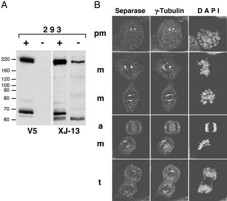
Figure 3.
Detection of endogenous human separase: monoclonal antibodies. (A) Whole-cell extracts were prepared from control 293 cells (−) or the cells induced to express recombinant V5-tagged full-length separase (+). Identical panels were probed with either V5-tag-specific (V5) or separase-specific (XJ-13) mAbs. (B) Endogenous separase was detected in 293 cells by using XJ-13 mAb and a TRITC-labeled secondary Ab, γ-tubulin was stained by polyclonal Ab, and Cy2-labeled secondary Ab and DNA was visualized by 4′,6-diamidino-2-phenylindole staining. pm, m, a, and t, prometaphase, metaphase, anaphase, and telophase cells, respectively.

