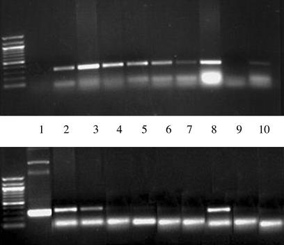
Figure 5.
Tissue localization of mole rat VEGF. DNA from various tissues was prepared as described in Methods. The presence or absence of mole rat DNA was ascertained by PCR with mole rat-specific primers, days 9–11 after operation. This procedure was repeated in three animals. (Upper) Presence of a specific band detecting 18S murine DNA sequences by PCR documenting the presence of DNA in each sample. (Lower) Presence or absence of mole rat VEGF in different tissues. Lanes: 1, control vector; 2, left (ischemic) lower proximal limb; 3, left lower distal limb; 4, right (control) lower proximal limb; 5, right lower limb; 6, left upper limb; 7, right upper limb; 8, liver; 9, negative control; 10, heart. Mole rat sequences were detected only in the proximal and distal portions of the ischemic limb and from the liver.

