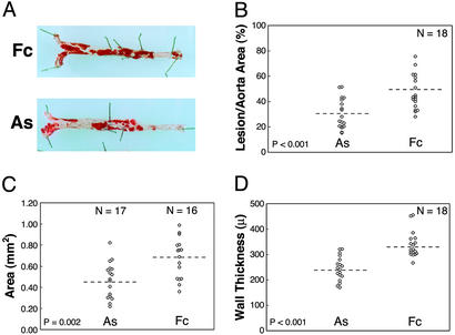
Figure 3.
AS treatment inhibits lesion progression. (A) Sudan IV-stained aortas isolated from animals representing the median lesion severity for the control (Fc) and AS groups. Atherosclerotic involvement for each mouse was determined by measuring the percent area of Sudan IV+ lesions in the aorta (B), the transverse area of lesions at the aortic sinus (C), and the wall thickness along the inner curve of the aortic arch (D). Statistical comparisons for the AS and Fc groups were based on the independent groups Student's t test. Results from two experiments (n = 18 total mice per group) were statistically significant when analyzed separately and in combination. Dashed lines indicate means.

