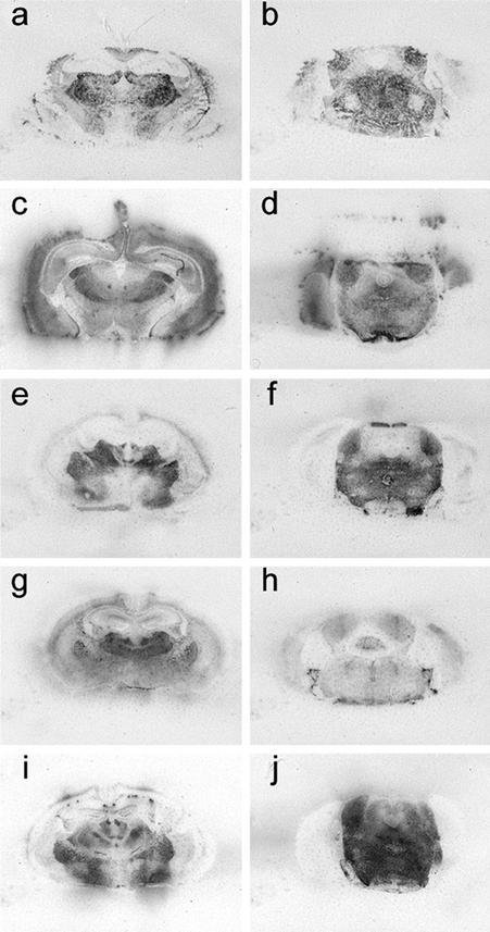
Figure 2.
PrPSc deposition shown by histoblotting in Tg22372 mice inoculated with different forms of CJD and FI. Coronal sections through the thalamic-hippocampal area (a, c, e, g, and i) or midbrain-pons area (b, d, f, h, and j) are depicted. Histoblots showing PrPSc deposition patterns after inoculation with sCJD(MM1) (RG) (a and b), fCJD(E200K) (c and d), FFI (e and f), sFI (g and h), and vCJD (i and j).

