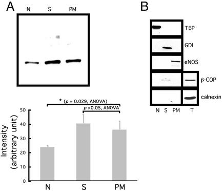
Figure 2.
Subcellular localization of ER46. (A) Immunoblot of ER46 among different organelles with F10 Ab. Organelles were isolated from 106 pretreated cells. Proteins were precipitated by ethanol from each fraction before use. N, nucleus; S, cytosol; PM, plasma membrane. Densitometric values were determined from three independent experiments and expressed as means ± SD, with P values of compared means as noted. (B) Examination of fraction purity by immunoblotting various protein markers from the N, S, PM, and total lysate (T). TBP, TATA-box binding protein; GDI, GDP dissociation inhibitor.

