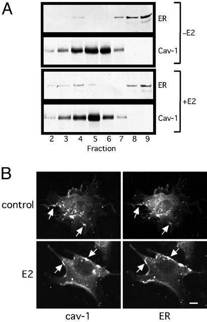
Figure 3.
Localization of membrane ER46 in caveolae. (A) Immunoblot of ER46 and cav-1 from isolated caveolae. Pretreated EA.hy926 cells were incubated with vehicle alone (−E2) or 30 nM E2 (+E2) for 10 min before fractionation. Fractions 2–9 were subjected to immunoblotting with anti-Cav-1 and F10 Abs. (B) Confocal microscopy of cell-surface cav-1 and ER46 with anti-Cav-1 and F10 Abs. Arrow, overlapped signals. (Bar = 10 μm.)

