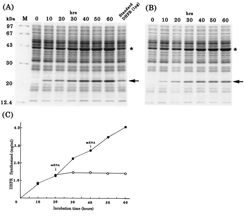
Figure 3.
Protein synthesis in the dialysis system. (A and B) Coomassie blue-stained SDS polyacrylamide gels showing DHFR synthesis with (A) or without (B) addition of new mRNA. Arrows and asterisks mark DHFR and creatine kinase, respectively. The standard sample was prepared by mixing a reaction mixture without mRNA with known amounts of DHFR before loading onto the gel. (C) Amounts of DHFR synthesized as determined from densitometric scans of the gels in A (closed circles) and B (open circles).

