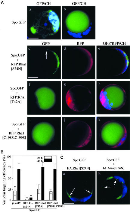
Figure 1.
Rha1[S24N] Strongly Inhibits the Vacuolar Trafficking of Spo:GFP.
(A) GFP patterns of Spo:GFP in the presence of various Rha1 mutants. Protoplasts were transformed with Spo:GFP alone or together with RFP:Rha1[S24N], RFP:Rha1[T42A], or RFP:Rha1[C198S,C199S], and the localization of GFP and RFP signals was examined. The GFP patterns shown here are representative of the protoplasts at 48 h after transformation. Blue indicates autofluorescent signals of chlorophyll. The arrow in (c) indicates the punctate stain. CH, chloroplasts. Bars = 20 μm.
(B) Quantification of the degree of inhibition. Protoplasts were transformed with the constructs indicated. pCaMV (for Cauliflower mosaic virus), a construct identical to RFP:Rha1 except for the region encoding the RFP:Rha1 fusion protein, was cotransformed with Spo:GFP as a balance for transformation and expression. Protoplasts with vacuolar patterns were counted from the whole population of transformed protoplasts at the times indicated. Three independent experiments were performed, and each time image from 50 randomly selected protoplasts was stored. The images were compared carefully based on GFP and chloroplast patterns to exclude the possibility of double scoring the same protoplasts. The patterns finally were scored to estimate the targeting efficiency. The error bars indicate standard deviations.
(C) GFP patterns of Spo:GFP in the presence of HA:Rha1[S24N] and HA:Ara7[S24N]. Protoplasts were transformed with the constructs indicated, and the localization of GFP signals was examined at 48 h after transformation. Arrows indicate punctate stains. Bar = 20 μm.

