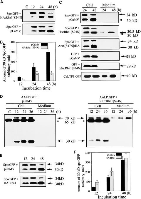
Figure 5.
Protein Gel Blot Analysis of Spo:GFP and AALP:GFP in the Presence of Rha1[S24N].
(A) Protein gel blot analysis of Spo:GFP in the presence of HA:Rha1[S24N]. Protoplasts were transformed with the constructs indicated. Protein extracts were prepared from transformed protoplasts at different times and used for protein gel blot analysis with anti-GFP antibody. pCaMV was used as a balance for transformation and expression. Proteins were prepared from protoplasts and the incubation medium separately. Equal amounts (20 μg) of total protein from protoplasts were analyzed by SDS-PAGE. Equal loading of total proteins was confirmed by Coomassie blue staining of protein blots after exposure. C, untransformed protoplasts.
(B) Quantification of the processed form of Spo:GFP. The intensity of the 30-kD band was measured at various times using image-analysis software. Identical experiments were performed three times to obtain means and standard deviations.
(C) Secretion of Spo:GFP into the medium. Protoplasts were transformed with the constructs indicated. Protein extracts were prepared from protoplasts and the medium at the times indicated and used in protein gel blot analysis with anti-GFP antibody. Equal amounts (20 μg) of total protein from protoplasts were analyzed by SDS-PAGE. In the case of protein in the medium, an equal volume was loaded. Equal loading of proteins was confirmed by Coomassie blue staining of protein blots after exposure.
(D) Protein gel blot analysis of AALP:GFP in the presence of RFP:Rha1[S24N]. Protoplasts were transformed with the constructs indicated. Protein extracts were prepared from the protoplasts and culture medium and used in protein gel blot analysis. The protein species indicated by asterisks at 65 kD is a cross-reactive endogenous protein species detected by the GFP antibody. Equal amounts (20 μg) of total protein from protoplasts were analyzed by SDS-PAGE. In the case of protein in the medium, an equal volume was loaded. Equal loading of proteins was confirmed by Coomassie blue staining of protein blots after exposure.

