
Figure 5.
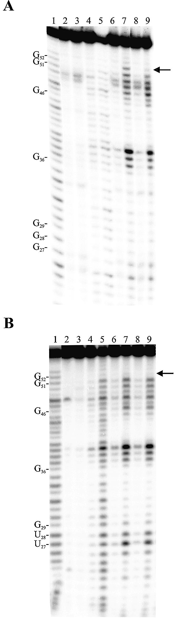
Typical autoradiograms of 10% PAGE of Mg2+-induced cleavage experiments. (A) The ribozyme possessing the original sequence (i.e. G27C12/G28C11). (B) The ribozyme possessing a P1.1 stem mutated to U27U12/U28U11. The 5′ end-labeled ribozymes were incubated in the presence of substrate, but in the absence of Mg2+ (lanes 2 and 3), or in the absence of substrate, but in the presence of Mg2+ (lanes 4 and 5). The remaining lanes contain the ribozymes, Mg2+ and either the substrate (lanes 6 and 7) or the 3′ product (lanes 8 and 9). Lanes 2, 4, 6 and 8 are incubations of 15 min, while lanes 3, 5, 7 and 9 are incubations of 120 min. Alkaline hydrolysis (lane 1) and RNase T1 hydrolysis (data not shown) of the 5′ end-labeled ribozymes were performed in order to determine the location of the metal ion-induced cleavage products. The locations of the RNase T1 major cuts are indicated on the left. The arrow on the right indicates the location of the G52.