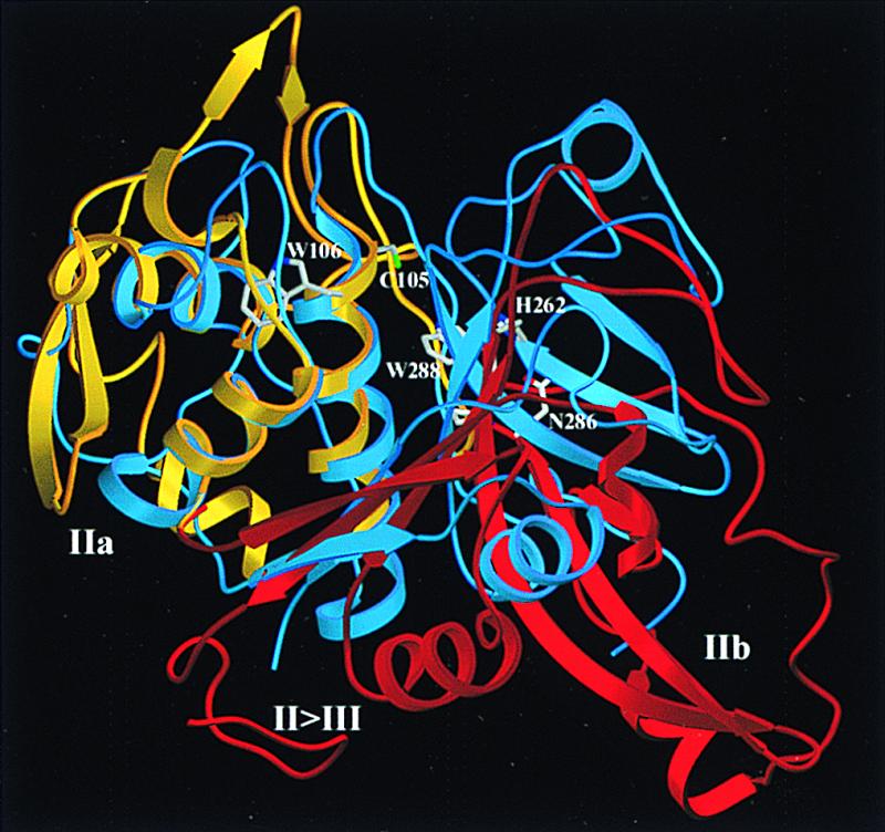
Figure 3.

Superposition of the m-calpain catalytic domain and papain. The papain-like part of the catalytic domain (gold, dIIa) and the barrel-like subdomain dIIb (red) are superimposed with papain (18) (blue) after optimal fit of the left-side papain half to the helical subdomain dIIa. The active site residues Cys105L, His262L, and Asn286L, and Pro287L, Trp288L, and Trp106L are shown in full structure. This “standard view” of papain-like cysteine proteinases (18) is obtained from Fig. 1 by a 90° rotation around a horizontal axis. The figure was made with setor (34).
