Figure 3.
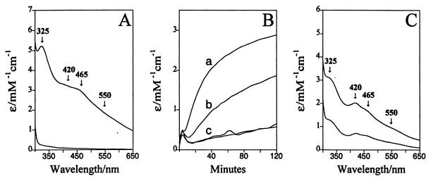
NifS-dependent in vitro Fe-S cluster assembly. (A) UV-visible spectrum of NifU-1 before in vitro Fe-S cluster assembly [before l-cysteine was added to initiate assembly (lower spectrum) and after 140 min of in vitro cluster assembly (upper spectrum)]. The postassembly spectrum shown in A is the maximum that could be obtained. (C) UV-visible spectrum of NifU-1(Asp37Ala) before in vitro cluster assembly (lower spectrum) and after 80 min of in vitro Fe-S cluster assembly (upper spectrum). The postassembly spectrum shown in C represents approximately 60% of the maximum that could be obtained. (B) Time dependence of Fe-S cluster assembly as monitored by the change in extinction coefficient at 465 nm vs. time after initiation of the Fe-S cluster assembly reaction. The time dependence for cluster assembly shown in line a of panel B corresponds to the same sample shown in panel A. Line b of panel B shows cluster assembly under the same conditions as for line a, except that half of the amount of NifS was added to the assembly cocktail. Data shown in the lines labeled c are controls. One data set corresponds to conditions that are the same as for line a except that an altered form of NifS having the active-site Cys325 residue substituted by alanine was used. The other data set in C corresponds to conditions that are the same as used for line a, except that an altered form of NifU-1 having the Cys62 residue substituted for by alanine was used. Conditions for Fe-S cluster assembly are described in Materials and Methods.

