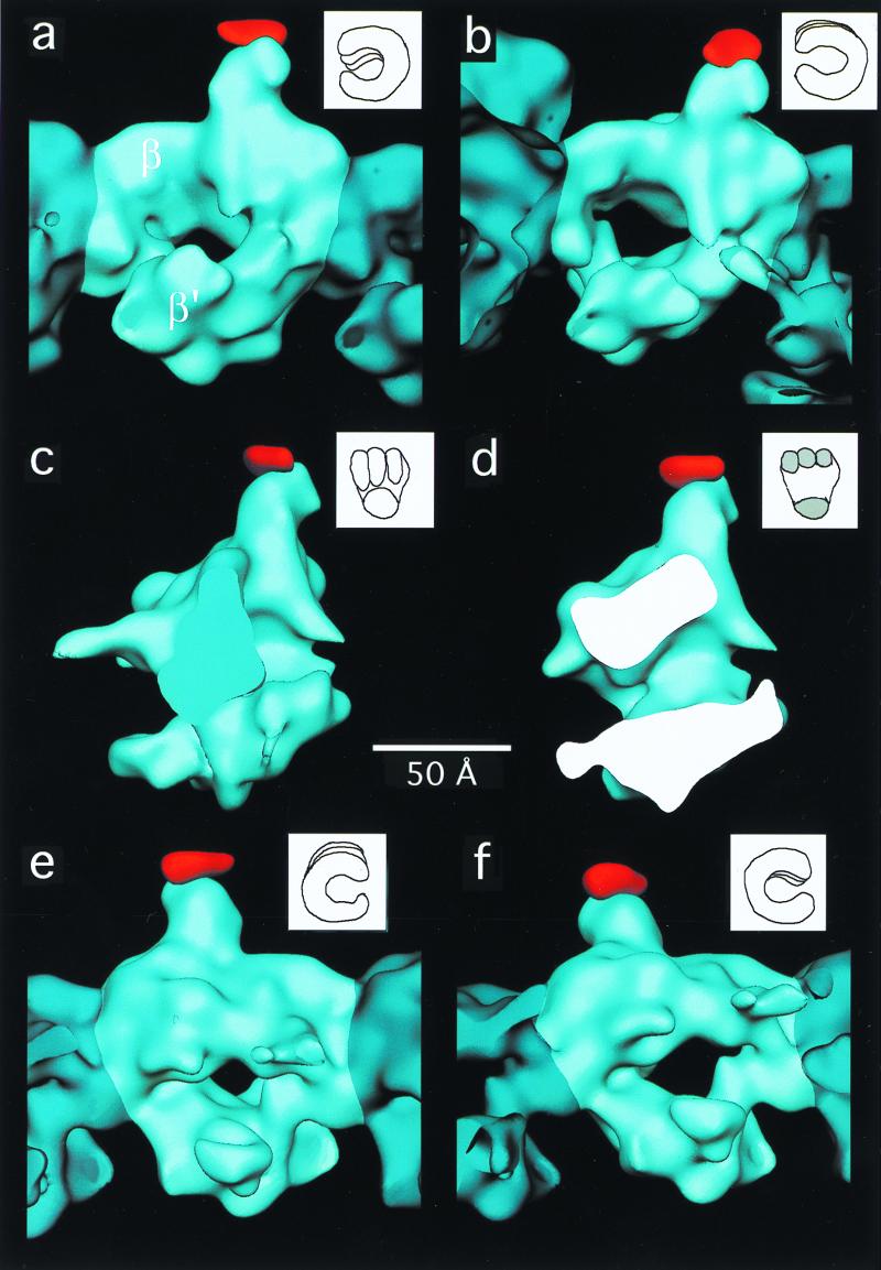
Figure 4.
Views of the three-dimensional reconstruction of wild-type E. coli core RNAP (blue) with an extra density (red) corresponding to the SPA insertion in the β-subunit. In each view, a single core RNAP molecule is highlighted (light blue). For each view, a drawing of a “hand” is shown (Upper Right) in an orientation similar to that of the RNAP molecule. The difference map (red density) was calculated in real space. (a) View parallel to the helix axis. (b) View showing the wide end of the channel down its axis. (c) View perpendicular to the channel. (d) Cross section of the molecule in an orientation similar to the one shown in c. (e) View corresponding to the opposite side shown in a. (f) View showing the narrow end of the channel down its axis.

