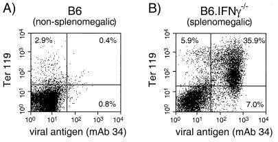
FIG. 7.
Flow cytometric analysis of FV-infected spleen cells from a persistently infected B6 mouse and a splenomegalic B6.IFN-γ−/− mouse at 18 weeks postinfection. Live nucleated spleen cells were stained for cell surface expression of FV glycosylated Gag protein (MAb 34) and Ter 119, an erythroid precursor cell marker. (A) The spleen from a representative persistently infected B6 mouse (nonsplenomegalic) had normal percentages of erythroid progenitor cells with very little infection. Lymphocyte and monocyte subset levels were also normal (data not shown). (B) The spleen from a representative splenomegalic B6.IFN-γ−/− mouse contained a predominance of infected erythroid progenitor cells. Ten different splenomegalic IFN-γ-deficient mice were analyzed and had levels of FV infection in erythroid precursor cells (Ter 119+) ranging from 30 to 80% of the spleen cells (data not shown).

