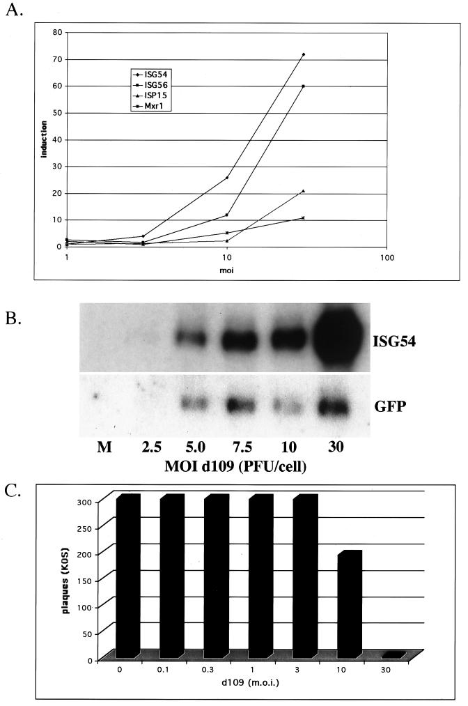
FIG. 2.
Induction of interferon-stimulated genes and antiviral effect as a function of MOI. (A) Microarray analysis was performed comparing mock-infected cells and cells infected with d109 at the indicated MOI as described in Materials and Methods. Shown are the induction ratios as a function of MOI for the four most highly induced interferon response genes. (B) Abundance of IFI54 and GFP in cells infected with d109 at various MOI was examined by Northern blot analysis. Total mRNA was isolated from mock-infected cells and cells infected with the indicated MOI of d109 at 24 h postinfection. (C) The antiviral effect of d109 is shown as a function of MOI. Monolayers of HEL cells were infected with the indicated MOI of d109 and 24 h later were used in a plaque assay with ≈300 PFU of KOS. Shown are the resulting numbers of plaques that developed 3 days later.

