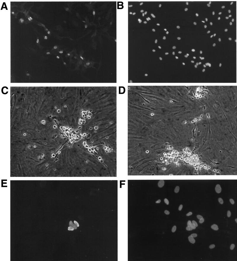
FIG. 4.
KSHV lytic induction and plaque formation in TIME cells treated with TPA. (A) Anti-ORF 59 immunofluorescence of KSHV-infected TIME cells induced for 3 days with TPA. (B) DAPI stain of panel A. (C and D) Light microscope pictures of infected TIME cells (1% LANA positive) 5 days after TPA induction. (E) Anti-ORF 59 immunofluorescence of infected TIME cells 3 days after TPA induction. (F) DAPI staining of panel E.

