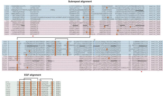
Figure 1.

Multiple sequence alignment of reelin repeats. Alignments of eight subrepeat A (cyan background) and eight subrepeat B (pink background) are shown at the top, and the central EGF repeats (green background) are shown at the bottom. β-strands in the repeat 3 are denoted with black underlining. The red dots under the alignment indicate the positions of residues making direct side-chain coordination to Ca2+. Cysteine residues are highlighted in orange and defined disulfide pairings are bracketed. Conserved arginine in subrepeat A (blue) and aspartate in EGF (red) implicated in the inter-subdomain contact are marked by circles at the top of the alignment.
