Figure 2.

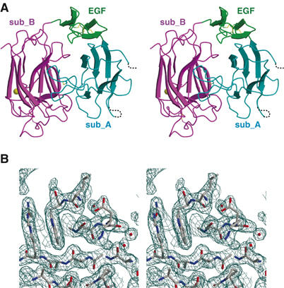
Structure of reelin repeat 3. (A) Stereo presentation of repeat 3. Subdomains are differently colored: subrepeat A (cyan), EGF (green), and subrepeat B (magenta). Bound calcium ion and disulfide bridges are shown as a gold sphere and yellow stick model, respectively. In subrepeat A, segments missing in the final model owing to poor electron density are indicated by dotted lines. (B) Stereo diagram of weighted 2∣F0∣−∣FC∣ electron density map in the region corresponding to the Asp-box motif. Figures were prepared with MOLSCRIPT (Kraulis, 1991), CONSCRIPT (Lawrence and Bourke, 2000), and RASTER3D (Merritt and Bacon, 1997).
