Figure 5.

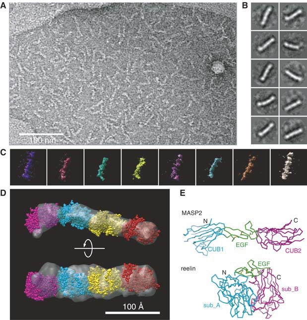
Structure of four-domain reelin fragment. (A) Representative raw image of the electron micrograph of the negatively stained R3–6 fragment. (B) 2-D class averages obtained from the untilted electron micrographs. Ten most highly populated classes, each derived from 25 to 46 particles, are shown. The width of each panel corresponds to 376 Å. (C) A gallery of typical 3-D electron tomograms obtained from individual R3–6 particles. (D) Predicted domain organization within the signaling-competent R3–6 fragment. 3-D structure of the four-domain fragment was created from the 10 most similar tomograms by 3-D averaging and presented as a molecular envelope (gray). Four complete space-filling models for the reelin repeat 3 (magenta, cyan, yellow, and red) are fitted into the envelope. (E) Difference between MASP2 and reelin in the subdomain arrangement. The complete model of the single reelin repeat (bottom) was superposed with a three-subdomain structure of MASP-2 (top, 1NT0) at their EGF module and is presented in the same orientation. Subdomains are differently color-coded and N- and C-termini are labeled.
