
Figure 2.
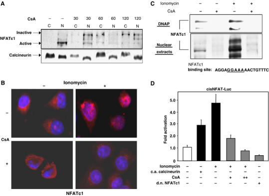
The Ca2+/calcineurin signaling pathway is active in pancreatic cancer cells. (A) Immunoblot analysis of subcellular localization of NFATc1 in pancreatic cancer cells. Panc-1 cells were either left untreated or treated with 1 μM CsA in the presence of serum for the indicated time intervals. Following subcellular fractionation, nuclear and cytoplasmic fractions were analyzed separately for the presence of NFATc1 protein by immunoblotting. (B) Immunocytological detection of NFATc1 localization in Panc-1 cells. Cells were kept in the absence of serum and either left untreated or treated with 1 μM CsA for 60 min, 1 μM ionomycin for 30 min, or a combination of CsA treatment followed by ionomycin treatment. Ionomycin stimulation led to a rapid and efficient translocation of NFATc1 (red signals) from the cytosol to the nucleus, which was completely blocked by CsA pretreatment. (C) DNA pulldown assays using oligonucleotides representing the consensus NFAT-binding sequence. Panc-1 cells were either left untreated or treated with 1 μM CsA for 30 min, 1 μM ionomycin for 15 min, or a combination of CsA treatment followed by ionomycin treatment. DNA–protein complexes from nuclear extracts were collected by precipitation with streptavidin–agarose beads and analyzed by Western blotting. Both, nuclear translocation (lower panel) as well as DNA-binding activity (upper panel) of NFATc1, were significantly enhanced by ionomycin, but completely blocked by CsA treatment. DNAP=DNA pulldown. (D) Activities of an NFAT-responsive reporter gene construct in Panc-1 cells. Cells were cotransfected with constitutively active (c.a.) calcineurin or dominant negative (d.n.) NFATc1 expression constructs and/or treated with 1 μM CsA or 1 μM ionomycin as indicated. Reporter gene activities are expressed as ‘fold activation' relative to untreated controls.