
Figure 2.
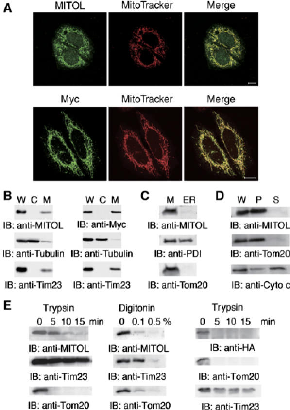
Specific localization of MITOL in the outer mitochondrial membrane. (A) Colocalization of MITOL with mitochondria. (Upper panels) HeLa cells were immunostained with anti-MITOL (green) antibody and MitoTracker (red). (Lower panels) HeLa cells expressing Myc-tagged MITOL were immunostained with anti-Myc antibody (green) and MitoTracker (red). (B) Subcellular fractionation indicates the localization of MITOL in mitochondria. Whole lysates (W) and cytosolic (C) and mitochondrial fractions (M) isolated from HeLa (left) or HeLa cells expressing Myc-MITOL (right) were immunoblotted with anti-MITOL, anti-Myc, anti-tubulin or anti-Tim23 mitochondria marker antibodies. (C) MITOL is not localized in ER. The mitochondrial fraction (M) and the ER-rich fraction (ER) isolated from HeLa cells were immunoblotted with anti-MITOL, anti-PDI (ER marker) or anti-Tom20 (mitochondria marker) antibodies. (D) Carbonate extraction assay indicates that MITOL is an integral membrane protein. Whole lysates (W), supernatant (S; peripheral protein) and pellet (P; integral protein) fractions after carbonate extraction from isolated mitochondria were immunoblotted with anti-MITOL, anti-Tom20 or anti-cytochrome c antibodies. (E) Sensitivity of MITOL to trypsin or digitonin treatment indicates MITOL localization in the mitochondrial outer membrane. Purified mitochondria from HeLa cells (left and middle panels) or cells expressing an N-terminally HA-tagged MITOL (right panel) were treated with trypsin or digitonin, and disappearance of MITOL along with mitochondrial markers was monitored by immunoblotting with anti-MITOL, anti-HA, anti-Tim23 or anti-Tom20 antibodies.