
Figure 3.
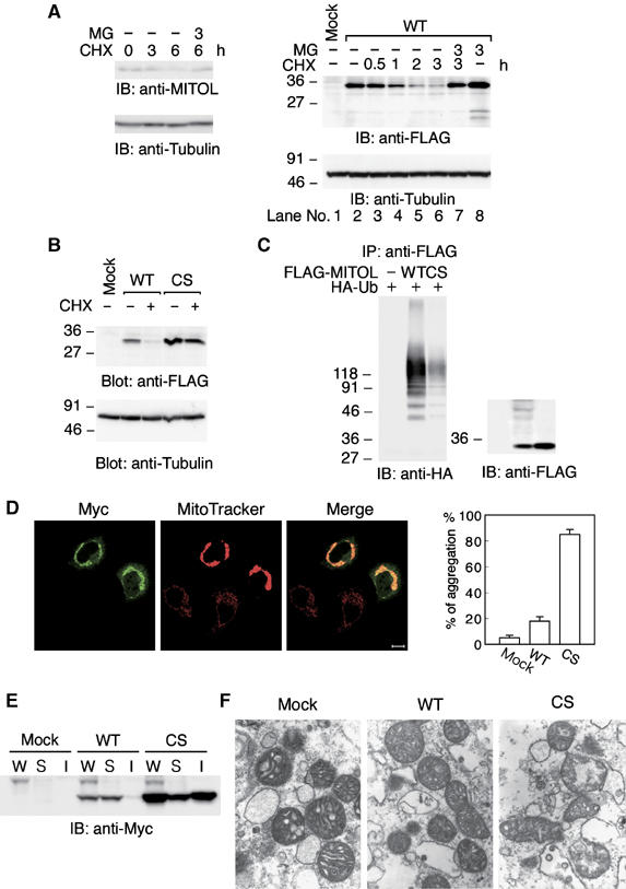
Rapid degradation of MITOL by autoubiquitination activity. (A) Effect of protein synthesis inhibitor CHX on MITOL protein expression. HeLa cells (left panel) or HeLa cells expressing FLAG-MITOL WT (right panel) were treated with or without CHX (10 μg/ml) for indicated times in the presence or absence of MG132 (50 μM) and cell lysates were immunoblotted with anti-MITOL, anti-FLAG or anti-tubulin antibodies. (B) No degradation of MITOL CS mutant. HeLa cells or HeLa cells expressing FLAG-MITOL WT or FLAG-MITOL CS mutant were treated with or without CHX (10 μg/ml) for 3 h and immunoblotted with anti-FLAG or anti-tubulin antibodies. (C) Polyubiquitination of MITOL WT but not MITOL CS mutant. Lysates of HeLa cells cotransfected with control vector, FLAG-MITOL WT or FLAG-MITOL CS mutant with HA-tagged ubiquitin (HA-Ub) were immunoprecipitated with anti-FLAG antibody agarose beads and immunoblotted with anti-HA or anti-FLAG antibodies. (D) Mitochondrial aggregation by MITOL CS mutant. HeLa cells expressing Myc-MITOL WT and CS mutant were stained with anti-Myc antibody and MitoTracker. Bar, 10 μm. Mitochondrial aggregation induced by MITOL WT or CS mutant was measured by counting at least 100 cells (right panel). Error bars represent s.d. n=3. (E) Accumulation of MITOL CS mutant in the insoluble fraction. Whole lysates (W) and soluble (S) and insoluble (I) fractions were isolated from HeLa cells or HeLa cells expressing Myc-MITOL WT or CS mutants and immunoblotted with anti-Myc antibody. Bars, 10 μm. (F) Electron microscopic analysis indicated severe mitochondrial damage in MITOL CS mutant-overexpressing cells. Mitochondrial morphology in control vector, MITOL WT or CS mutant-transfected cells was examined by electron microscopic analysis at 27 600-fold magnification.