
Figure 5.
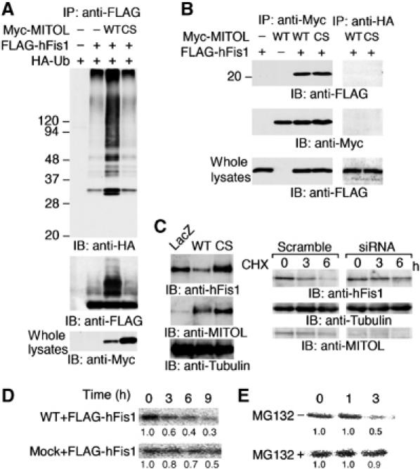
MITOL associates with and ubiquitinates mitochondrial fission protein hFis1. (A) Ubiquitination of hFis1 by MITOL. Lysates of HeLa cells transfected with indicated vectors were immunoprecipitated with anti-FLAG antibody and immunoprecipitates were immunoblotted with anti-HA or anti-FLAG antibodies. Whole lysates were immunoblotted with anti-Myc antibody. (B) Interaction of MITOL with hFis1. Lysates of HeLa cells transfected with indicated vectors were immunoprecipitated with anti-Myc antibody and immunoprecipitates were immunoblotted with anti-FLAG or anti-Myc antibodies. To demonstrate the specificity of the co-immunoprecipitation, anti-HA antibody was used as a negative control. Whole lysates were immunoblotted with anti-FLAG antibody to confirm the expression of FLAG-hFis1. (C) Accumulation of endogenous hFis1 by MITOL dysfunction. Lysates of HeLa cells infected with indicated adenovirus vectors were immunoblotted with anti-hFis1, anti-MITOL or anti-tubulin antibodies (left panel). Lysates of HeLa cells transfected with scramble or MITOL siRNA1 were treated with CHX (10 μg/ml) for indicated times and immunoblotted with anti-hFis1, anti-tubulin or anti-MITOL antibodies (right panel). (D) MITOL expression caused a rapid turnover of hFis1. Pulse–chase experiment was performed. HeLa cells expressing FLAG-hFis1 or FLAG-hFis1/MITOL WT were labeled for 60 min with [35S]Met/Cys labeling mixture and chased in complete DMEM for indicated periods. (E) Effect of MG132 on hFis1 turnover promoted by MITOL overexpression. HeLa cells expressing FLAG-hFis1/MITOL WT in the absence or presence of MG132 were similarly chased for indicated periods. Cells were lysed and immunoprecipitated with anti-FLAG antibody and immunoprecipitated FLAG-hFis1 was visualized by autoradiography. The result (D or E) is representative of three independent experiments. Relative values estimated by NIH image analysis are indicated below.