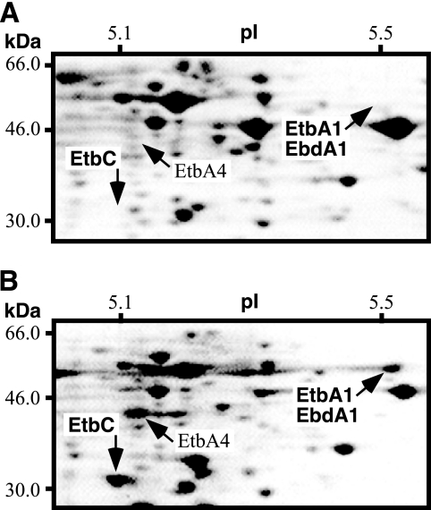
FIG. 2.
Two-dimensional polyacrylamide gel electrophoresis analysis of soluble proteins from RHA1 cells before (A) and after (B) incubation in the presence of biphenyl for 16 h. Proteins were separated in an isoelectric point (pI) gradient from 4 to 7 and then in a 12% polyacrylamide gel, after which they were stained with Coomassie brilliant blue R250. The area of the pI gradient from 5.1 to 5.5 and of molecular mass from 30.0 to 66.0 kDa is shown. The pI and molecular mass scales are indicated at the top and left of each panel, respectively.

