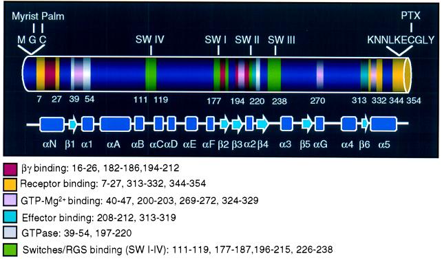
Figure 4.
Primary and secondary structures of Gαi1. The primary and secondary structure as well as functional domains of Gαi1 (12, 28), which are similar to that of Gαi3, are indicated in the diagram. The N terminus (amino acids 1–27) is involved in membrane anchoring (myristoylation and palmitoylation) (35), as well as receptor (11, 12, 26) and βγ (12) binding. The C terminus has been shown to participate in binding to effectors (13) and GDP/GTP (31, 32) as well as to receptors (11, 12, 26). The switch regions (I–III) constitute the binding site for RGS proteins (RGS 4) (41) and adenylyl cyclase (13), as well as βγ subunits (12).

