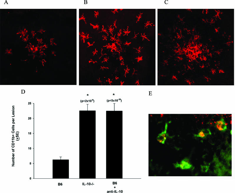
Figure 2. Cellular Infiltrates in CNV Lesions.
(A–C) On day 7 following laser treatment, whole mount stains were performed to determine the cells present in the area of the neovascular complex. Stains for CD11b were performed on (A) B6, (B) IL-10 −/−, and (C) B6 mice treated with neutralizing anti-IL-10. Images were take by confocal microscopy (magnification 200×) centered on the laser lesion.
(D) The number of CD11b+ cells per lesion was counted (200× high-power field centered on the laser lesion) (B6, 6.3 ± 0.9; IL10 −/−, 22.6 ± 2.1; B6 + anti-IL10, 22.5 ± 2.5). Asterisks indicate values significantly different from control.
(E) Dual staining was performed on day 7 following laser treatment using FITC-conjugated anti-CD11b (green) and PE-conjugated anti-F4/80 (red) (magnification 400×).

