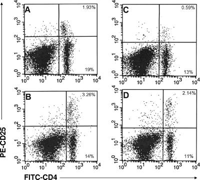
FIG. 4.
Numbers of CD4+ CD25+ T cells in the inguinal lymph nodes of Borrelia-vaccinated and -challenged mice without (A and B) and with (C and D) treatment with anti-CD25 antibody at days 8 (A and C) and 20 (B and D) after infection. The upper right quadrants show the percentages of CD4+ CD25+ T cells. The lower right quadrants show the percentages of CD4+ CD25− T cells.

