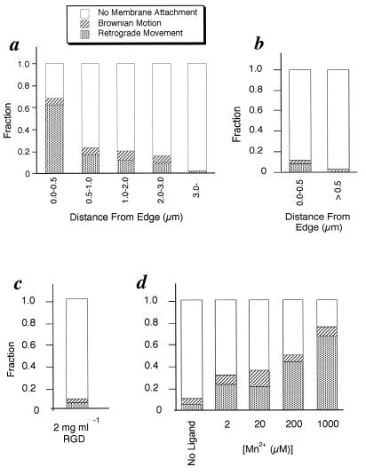
Figure 2.
Histograms are shown of the probability of bead binding. (a) The position dependence of beads coated with a low concentration of FNIII7–10 (total n = 272). At this concentration, we estimate that 300 FNIII7–10 molecules are bound per bead (4–10/bead-membrane contact area). (b) The control experiment for a. Beads were coated with 100% BSA (total n = 75) instead of FNIII7–10. (c) The same experiment as a in the presence of 2 mg/ml RGD peptide, which inhibits fibronectin binding to the β1 integrin (19, 20). This experiment was done within 0.5 μm from the edge (total n = 94). This result coincides with ref. 1. (d) Edge binding of FN beads is measured as a function of [Mn2+], which is reported to change the affinity of integrin for fibronectin (20–22). The binding probability is dependent on the concentration of Mn2+ (total n = 252).

