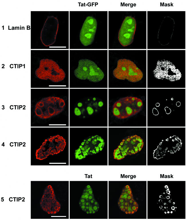
FIG. 5.
Localization of Tat-GFP and Tat in the presence of CTIP1 and CTIP2 in microglial cell nuclei. Cells were transfected with vectors expressing Tat-GFP (5 ng) in the absence (row 1) or the presence of HA-CTIP1 (row 2) and Flag-CTIP2 (rows 3 and 4). Cells were cotransfected with pCMV-Tat (500 ng) and Flag-CTIP2 (row 5). Cells were fixed 24 h after transfection, incubated with anti-lamin B, and stained with Alexa Fluor 568-conjugated secondary antibodies to detect endogenous lamin B and delineate the nucleus (row 1). To detect CTIP1 and CTIP2, cells were incubated with anti-HA (row 2) and anti-Flag (rows 3, 4, and 5) antibodies and stained with cyanine 3-conjugated secondary antibodies. To detect Tat, cells were incubated with monoclonal Tat antibodies and stained with cyanine 2-conjugated secondary antibodies (row 5). Masks were obtained after selection of the double-labeled pixels in the two-dimensional scatter histograms of gray values constructed from red and green images. Bars: 10 μm. HA-CTIP1 exhibited a diffuse staining pattern, with did not alter the Tat-GFP nucleolar and nuclear fluorescence (row 2). Flag-CTIP2 exhibited a ball-like staining in most of the nuclei. Tat-GFP and Tat were located within these ball-like structures that were distributed randomly in 80 to 90% of the nuclei (rows 3 and 5) or localized at the periphery of the inner nuclear membrane in 10 to 20% of nuclei (row 4). Tat-GFP and Tat colocalize with CTIP2 at the periphery of CTIP2-induced structures (columns 3 and 4).

