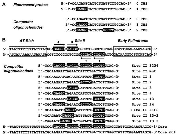
FIG. 2.
DNA duplexes used in this study. (A) Fluorescent probes and analogous competitor oligonucleotides. The sequences of the fluorescein (F)-labeled strand of the probe containing a single T antigen-binding site (TBS) and of the control probe are indicated. Similarly, the top strand of competitor oligonucleotides containing one, two, or no TBS is indicated. TBS are boxed in black. (B) Oligonucleotides derived form the SV40 origin. The sequence of the 64-bp double-stranded core origin is indicated. The AT-rich and early palindrome regions are boxed. The positions of site II and of the four pentanucleotide TBS are indicated. The bottom of the figure describes the sequence of the top strand of competitor oligonucleotides derived from the 31-bp site II or from the 64-bp core origin. Functional (not mutated) TBS are boxed in black and their numbers are specified in the nomenclature of each oligonucleotide. Site II oligonucleotides in which the spacer region between TBS 1 and 3 has been increased by 1, 2, or 3 bp are referred to as +1, +2, and +3, respectively.

